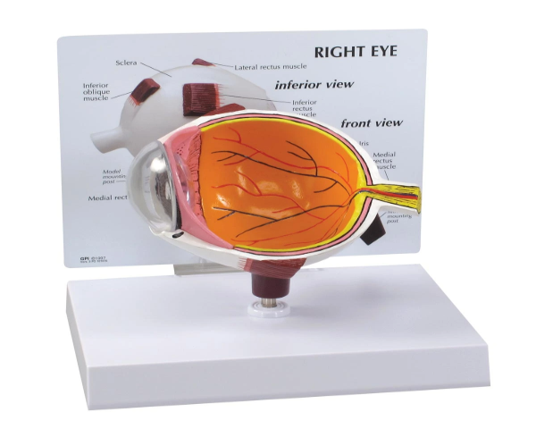
An Eye Anatomy Model is a magnified, three-dimensional representation of the human eye, designed to illustrate its intricate internal and external structures. These models are invaluable educational aids in ophthalmology clinics, medical schools, and biology classrooms. They typically feature dissectible components that allow users to explore the cornea, iris, lens, retina, optic nerve, and extraocular muscles. By providing a clear visual and tactile representation, the eye anatomy model helps students and patients understand complex conditions, surgical procedures, or the basic function of vision, making abstract concepts concrete and fostering a deeper appreciation for the complexity of the human eye.
 English
  Japan
Japan 


Reika Ogawa
視神経や水晶体などの構造も理解しやすいです。
Nathaniel Hughes
Good quality plastic and easy to clean.
Hiroki Arai
パーツの取り外しがしやすく、授業に使いやすいです。
Maya Long
Detailed and labeled perfectly—great for students.
Satoshi Inoue
細部までよく再現されていて、視覚の理解が深まりました。